
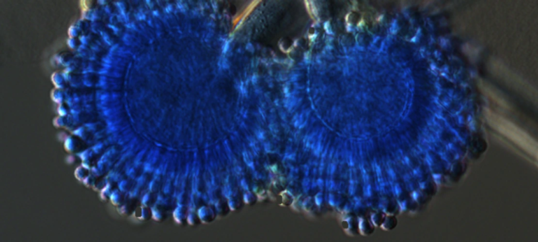
Aspergillus spore structure, lactophenol blue staining with Differential Interference Contrast (DIC) - Pummi Singh

Scanning Electron Microscope (SEM) image of the top of a Blue Papilio blumei butterfly wing. Brooke Massani

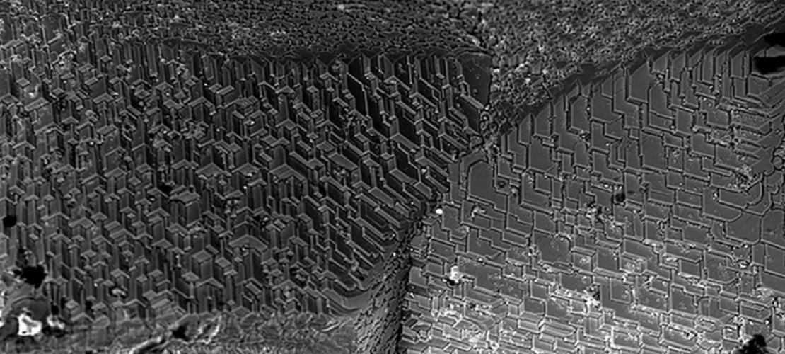
Focused Ion Beam (FIB) milled cross section in copper on the FEI Helios 660 G3 FIB-SEM DualBeam microscope. A protective cap of ion deposited platinum is necessary as polycrystalline copper mills at different rates which otherwise results in spires and curtains in the final cross section. (Dr. Paul Wallace)

Widefield and deconvolved image of fluorescently labeled fibroblasts - Patty Jansma

Drosophila neuropil - Transmission Electron Microscope (TEM) - Patty Jansma

Alveoli (lung) - Two color confocal microscopy, Green nuclei with red elastin. (R. Clark Lantz lab)
Geosciences Scanning Electron Microscope (Isabel Fay Barton)
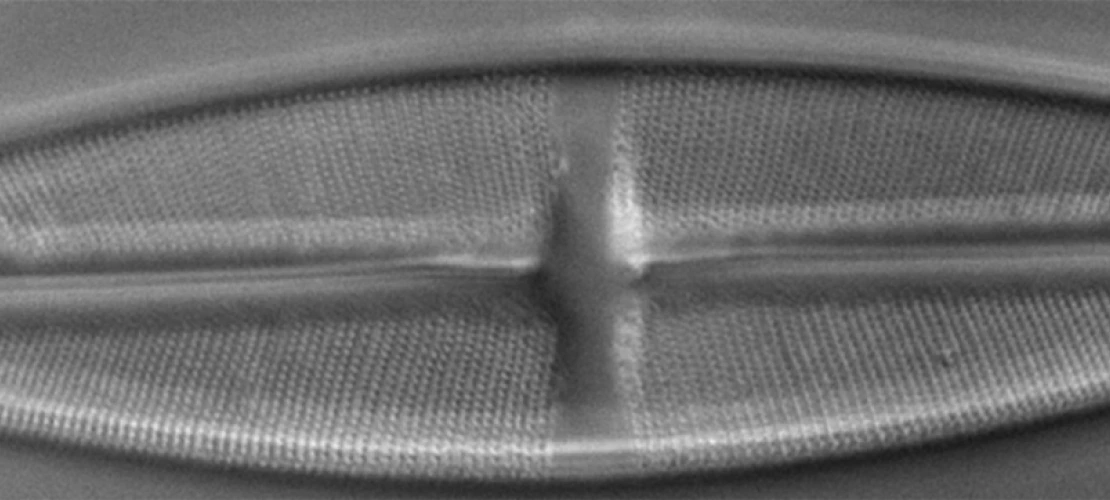
Differential Interference Microscopy (DIC/Nomarski) microscopy - unstained diatom at 40x, Zeiss LSM510 confocal, Doug Cromey

Luxol fast blue (histology stain for myelin) - Cromey & Grantham

Transmission Electron Microscope (TEM) Skeletal muscle with misplaced Z bands (Tony Day for Gregorio Lab)

Atomic Force Microscope (AFM) image of Perylene deposited on highly ordered pyrolytic graphite. (2 micron image) Derek Mangelsdorf (Armstrong Group) and Brooke Beam

Atomic Force Microscope (AFM) image of spin coated biodegradable polymer (polycaprolactone). (15 micron image) Jessi Gamboa (Yoon Group) and Brooke Massani

Geosciences by Scanning electron microscopy (SEM) - Isabel Fay Barton

Polarized light microscopy - picrosirious red stain of optic nerve (causes collagen to become birefringent) - Cromey & Grantham

Scanning Electron Microscope (SEM) image of the head of a fly (Hitachi S3400) - Kuiper Imaging, Juliane Weber, PhD

Scanning electron microscopy (SEM) image of the broken edge of mechanical pencil lead. Brooke Massani

Liver - Oil Red O stain for lipids (cryostat section) - Cromey
Scanning electron microscopy (SEM) image of the broken edge of mechanical pencil lead. Brooke Massani
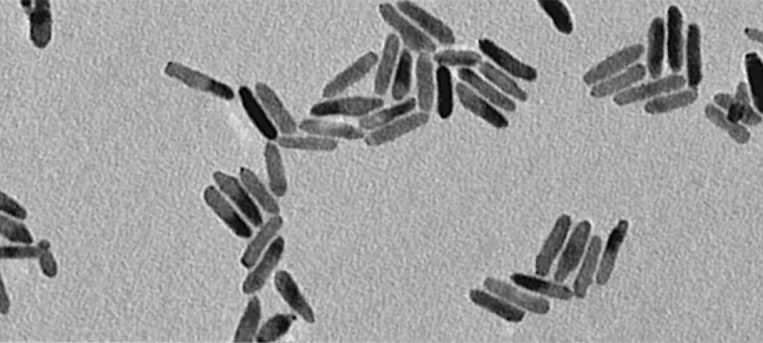
Transmission Electron Microscope (TEM) Metallic nano particles. Unstained (Nick Pavlopoulos – Pyun Lab)

Scanning electron microscopy (SEM) image of a circuit board. Dee Belle-Oudry

Transmission Electron Microscope (TEM) Multi-lamina lipid bilayers showing cross-hatch pattern. Ammonium molybdate negative stain (Tony Day)

https://wordart.com/
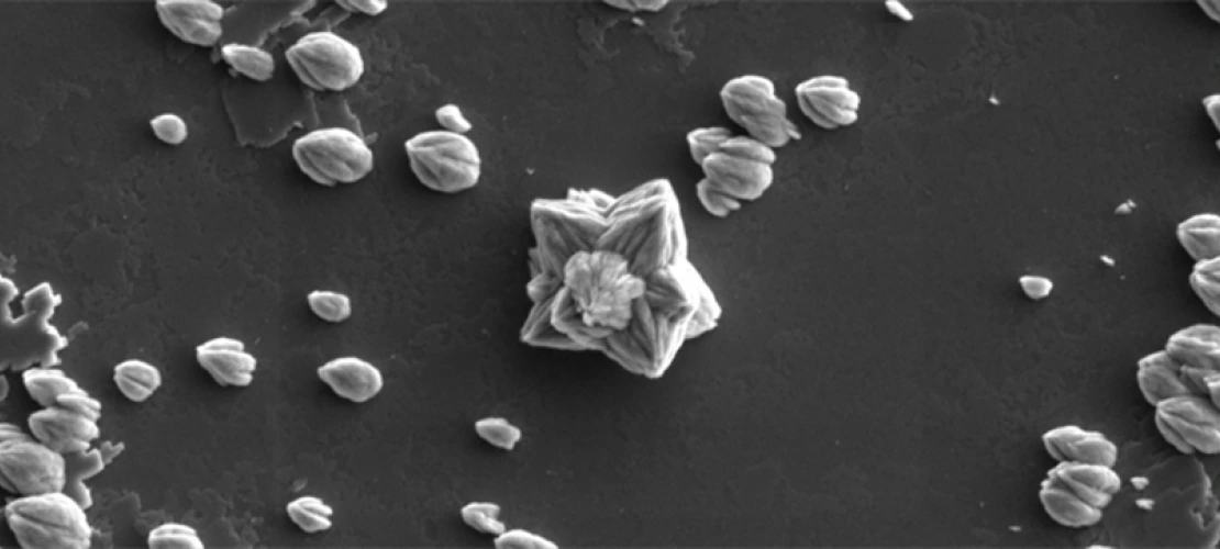
Scanning electron microscopy (SEM) image of Strontianite (SrCO3) crystals grown on a calcite surface. (FEI Helios 660) - Kuiper Imaging, Juliane Weber, PhD
University of Arizona microscopy-related News & Events

Did You Know That?
Featuring tips, tools, explanations, and information about things that are not quite microscopes, but might be of use to our users. Is there something you would like to see here? Contact us and let us know.
Full List | UA Resources | Non-UA Resources | Software/Computing | General Tips