-
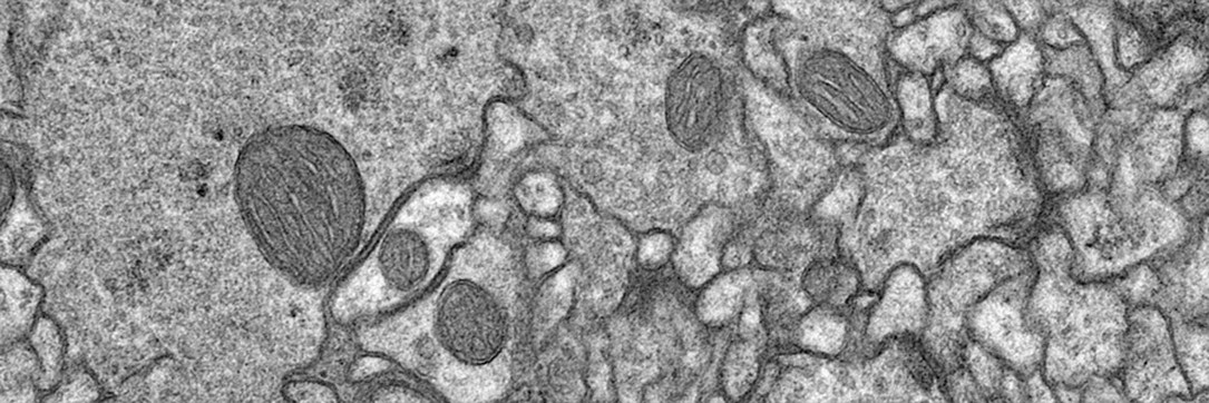
Transmission Electron Microscope (TEM) Skeletal muscle with misplaced Z bands (Tony Day for Gregorio Lab)
-
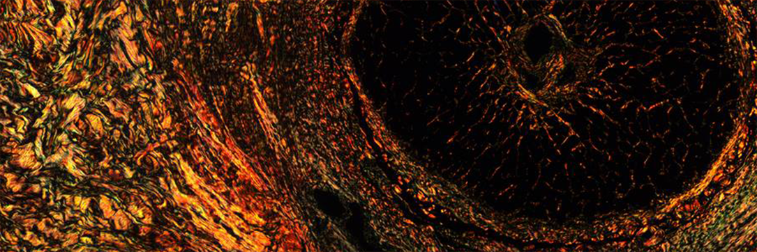
Polarized light microscopy - picrosirious red stain of optic nerve (causes collagen to become birefringent) - Cromey & Grantham
-
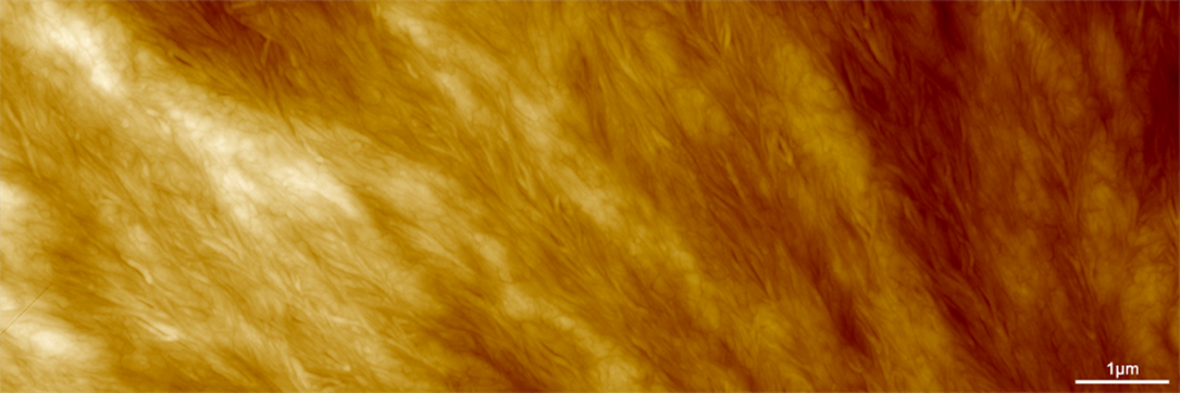
Atomic Force Microscope (AFM) image of spin coated biodegradable polymer (polycaprolactone). (15 micron image) Jessi Gamboa (Yoon Group) and Brooke Massani
-
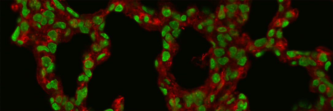
Alveoli (lung) - Two color confocal microscopy, Green nuclei with red elastin. (R. Clark Lantz lab)
-
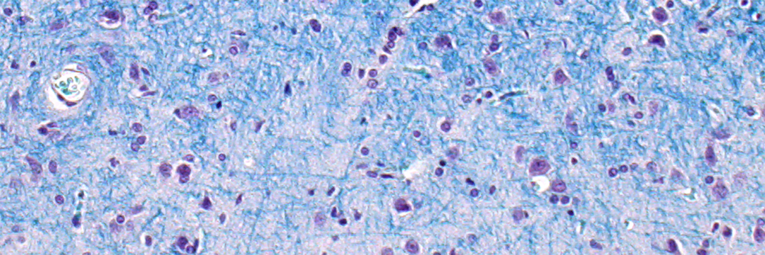
Luxol fast blue (histology stain for myelin) - Cromey & Grantham
-
Geosciences by Scanning electron microscopy (SEM) - Isabel Fay Barton
-
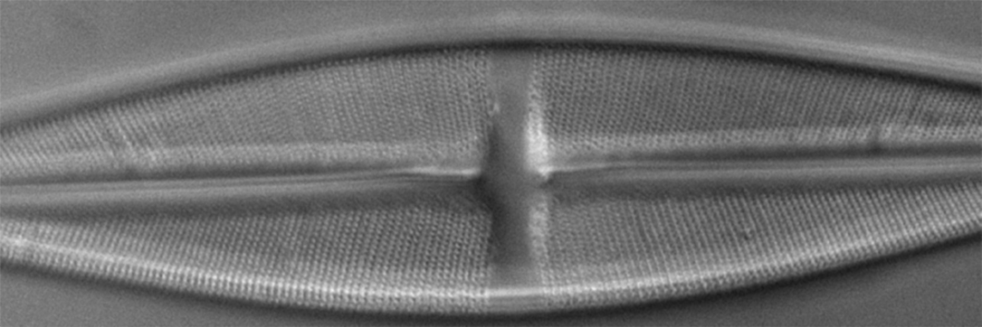
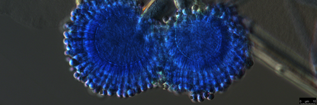
Differential Interference Microscopy (DIC/Nomarski) microscopy - unstained diatom at 40x, Zeiss LSM510 confocal, Doug Cromey
-
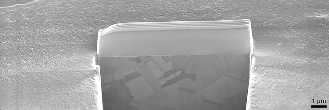
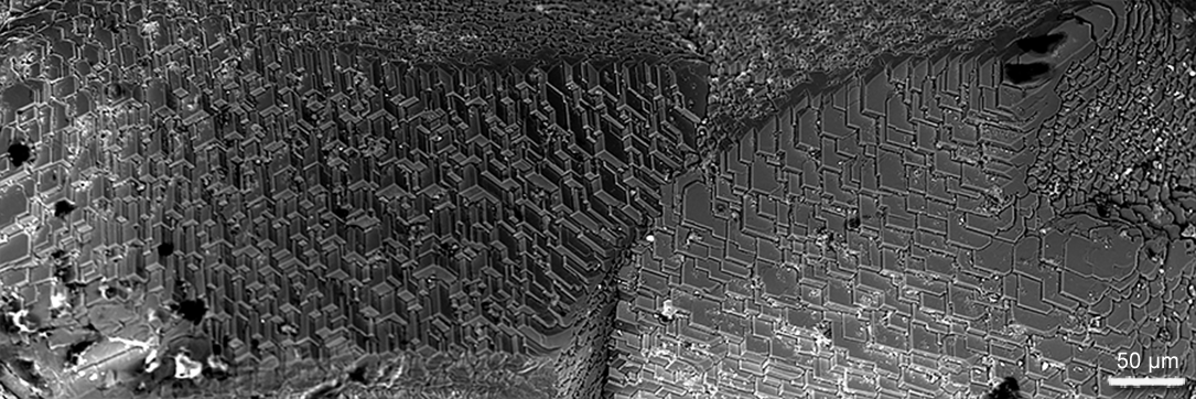
Focused Ion Beam (FIB) milled cross section in copper on the FEI Helios 660 G3 FIB-SEM DualBeam microscope. A protective cap of ion deposited platinum is necessary as polycrystalline copper mills at different rates which otherwise results in spires and curtains in the final cross section. (Dr. Paul Wallace)
-
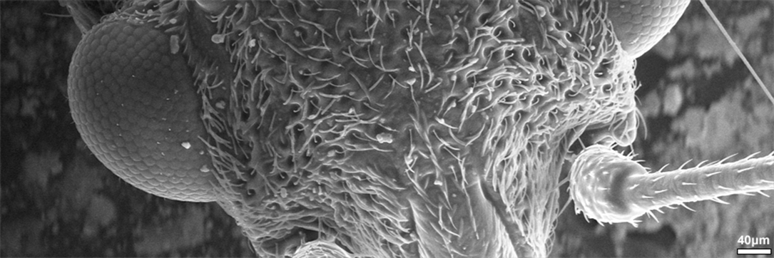
Scanning Electron Microscope (SEM) image of the head of a fly (Hitachi S3400) - Kuiper Imaging, Juliane Weber, PhD
-
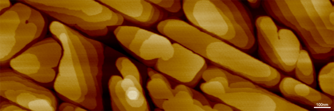
Atomic Force Microscope (AFM) image of Perylene deposited on highly ordered pyrolytic graphite. (2 micron image) Derek Mangelsdorf (Armstrong Group) and Brooke Beam
-
Drosophila neuropil - Transmission Electron Microscope (TEM) - Patty Jansma
-
Aspergillus spore structure, lactophenol blue staining with Differential Interference Contrast (DIC) - Pummi Singh
-
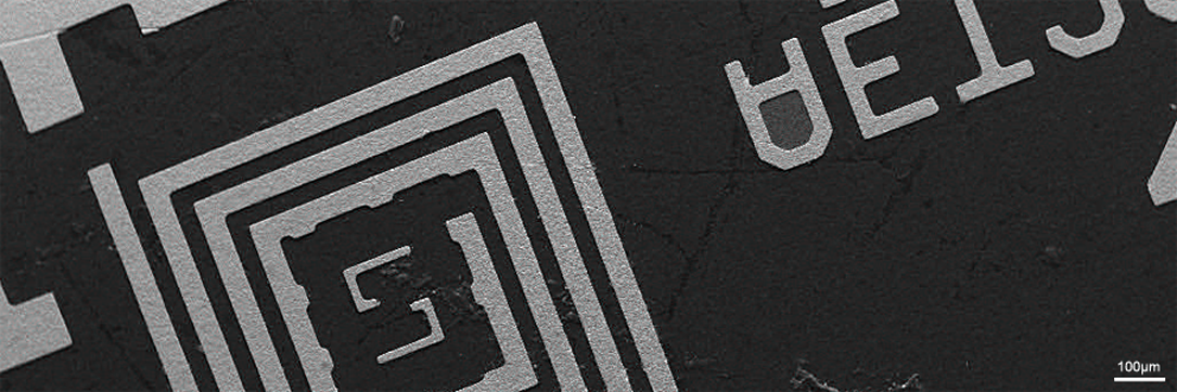
Scanning electron microscopy (SEM) image of a circuit board. Dee Belle-Oudry
-
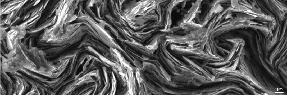
Scanning electron microscopy (SEM) image of the broken edge of mechanical pencil lead. Brooke Massani
-
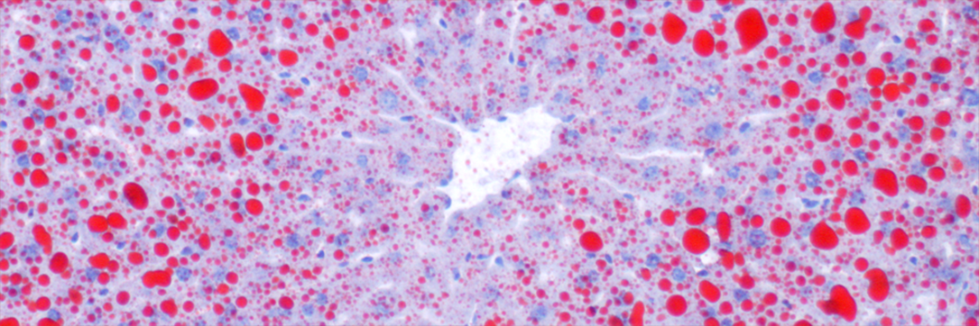
Liver - Oil Red O stain for lipids (cryostat section) - Cromey
-
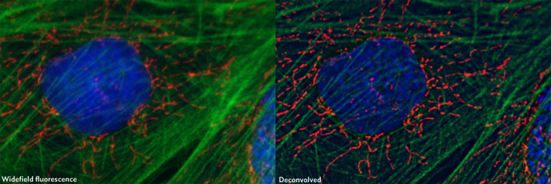
Widefield and deconvolved image of fluorescently labeled fibroblasts - Patty Jansma
-
Geosciences Scanning Electron Microscope (Isabel Fay Barton)
-
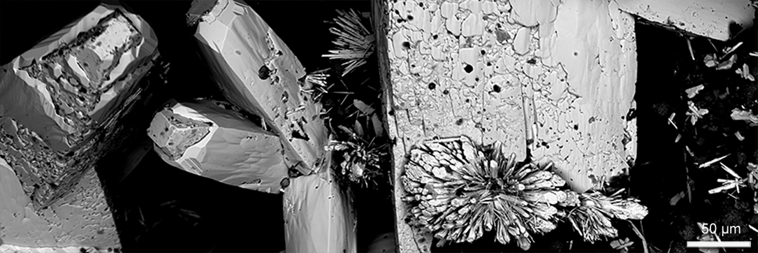
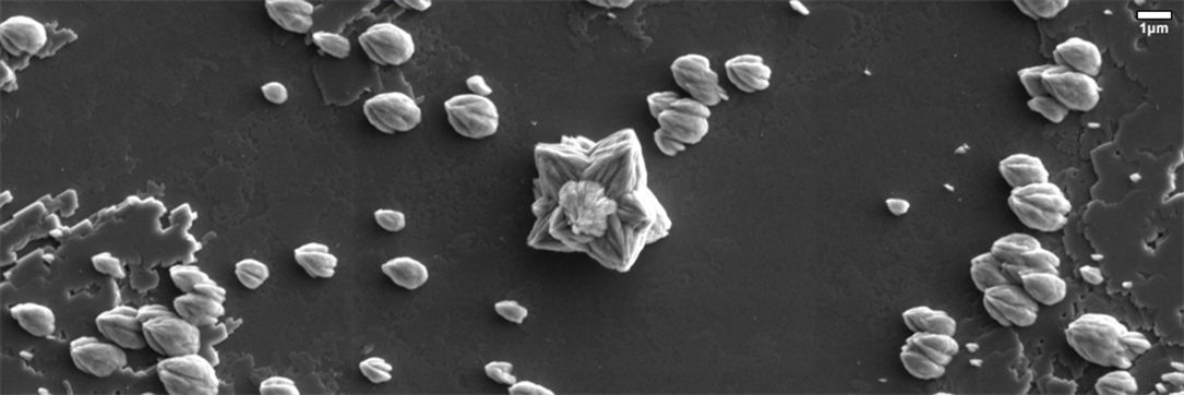
Scanning electron microscopy (SEM) image of Strontianite (SrCO3) crystals grown on a calcite surface. (FEI Helios 660) - Kuiper Imaging, Juliane Weber, PhD
-
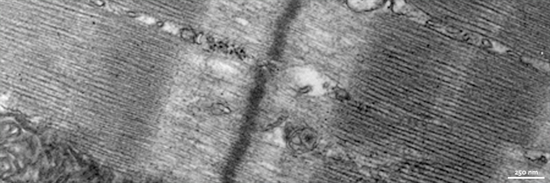
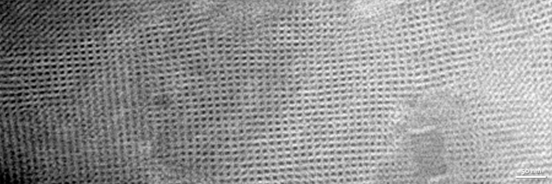
Transmission Electron Microscope (TEM) Multi-lamina lipid bilayers showing cross-hatch pattern. Ammonium molybdate negative stain (Tony Day)
-
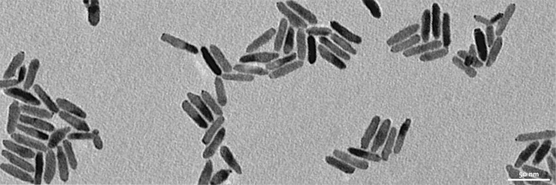
Transmission Electron Microscope (TEM) Metallic nano particles. Unstained (Nick Pavlopoulos – Pyun Lab)