
Introducing the Zeiss Axio Observer 7 inverted microscope with Apotome III
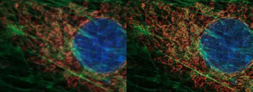
Fluocells 1 slide imaged using 63X oil objective. Left image was acquired using the Apotome and right image is the same data after software deconvolution. (BPAE cells with MitoTracker™ Red CMXRos, Alexa Fluor™ 488 Phalloidin, and DAPI)
Many of you may remember that about a year ago the ORP Imaging Cores - Optical hosted four different vendor’s on-site microscope demonstrations (Aug & Sept 2022, Microscopy Month). We used the information gathered from those demonstrations to successfully submit an internal Equipment Enhancement Fund proposal for this microscope (TRIF funds). Additional funds to complete the purchase came from the RII Core Facilities department budget.
Our goal was to have a microscope that could give confocal-like images of thinner samples that would be available at our Life Sciences North location on the UA Health Sciences campus.
Who would benefit?
- Users with samples such as cells on a coverslip, paraffin sections or standard frozen sections up to approximately 20um in thickness.
- The Apotome only works on fluorescent samples. The structured illumination used can provide confocal-like images and z-stacks.
- Users doing large area stitching of fluorescent samples will find this instrument to be faster than any of the other instruments in our core. Adding in the ability to capture z-stacks will be of benefit as well.
- For users wanting to add a near-IR fluorophore to their staining, this instrument can capture those wavelengths (in widefield mode only, no Apotome).
- The AI sample finder may be useful to labs with small un-stained samples that are hard to locate, with multiple sections on a slide that all need to be tile-scanned, and for imaging multiwell plates.
- People stitching standard histology slides should see a speed improvement and the AI sample finder can speed up the setup for tile scanning.
- The LED fluorescence illuminator offers some options for ex/em wavelengths that the Leica microscope is not as well suited for. The Axio Observer’s filters/excitation wavelengths can be seen on this FPbase webpage.
Weaknesses:
- The ZEN Blue software has quite a large number of options. This instrument might not be an appropriate starting point for a brand new “I’ve never used a high-end research microscope” user.
- Low contrast and/or dim fluorescent samples don’t work any better on the Apotome than they do on a standard confocal.
- Images acquired with the Apotome can be post-processed by software deconvolution and there is a noticeable improvement, with the caveat that not all samples will benefit from post-processing.
- This is a filter-based fluorescence microscope, so there may be instances where the spectral nature of the Marley point-scanning confocal would give better images.
- Because this instrument uses multi-band fluorescence filters, there may be some spectral bleedthrough that would be problematic for some research uses.
- Because the microscope’s objective nosepiece is already full, the 40X dry or the 100x oil objective are only available on request (in advance).
New user training will begin mid-August. If the interest is high, it may take some time for everyone to get trained.
The hourly rate for trained users is $25/hr. Contact Doug Cromey to arrange for user training ($82/hr, 2-3 sessions). The Axio Observer 7’s specifications.