
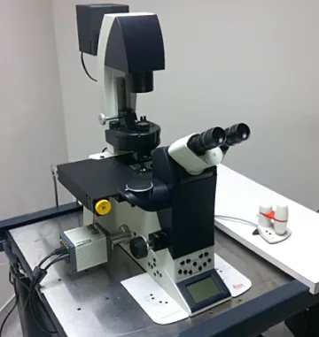
Leica DMI6000 motorized inverted microscope with two cameras
This mulitfunction fully-motorized inverted microscope can capture both 24bit color transmitted light images, and 16bit epi-fluorescence images.
The microscope includes a 5Mpix camera for color brightfield images (Leica DCF450), as well as in DIC (Nomarski), and Polarization [Note: crossed polars only, this is not a full-on POL microscope]. The sCMOS greyscale camera (Hamamatsu Flash 4.0) can capture 4Mpix images in 16bits at up to 30fps. The microscope has fluorescence cubes for dyes similar to DAPI, FITC/GFP, Rhodamine, and CY5 (to check your dyes with our filters, see our page at FPbase). The microscope can also do phase contrast imaging, but the optical path is not currently configured for capturing phase images. The microscope has 2.5x, 5x, 10x, 20x, 40x dry objectives and 40x, 63x, 100x oil objectives, as well as a 1.0/1.6x optivar for intermediate magnifications. The stage can accommodate microscope slides, multiwell plates*, and small culture dishes (35mm, 70mm)*. We have a BiopTechs Delta-T live cell culture dish controller to allow for long-term live cell imaging at 37 degrees C.
Capturing images is fairly easy and the Leica LAS-X software is able to allow users to stitch together multiple fields of view, creating a much larger image (color or greyscale cameras).
Our Resources page: http://microscopy.arizona.edu/ic-optical-resources
* low mag images through plastic dish bottoms are possible, but higher magnification image capture or DIC/POL images require the use of a coverslip thickness (#1.5, 0.170mm) glass bottom on the dish.